about us

HOME / ABOUT US

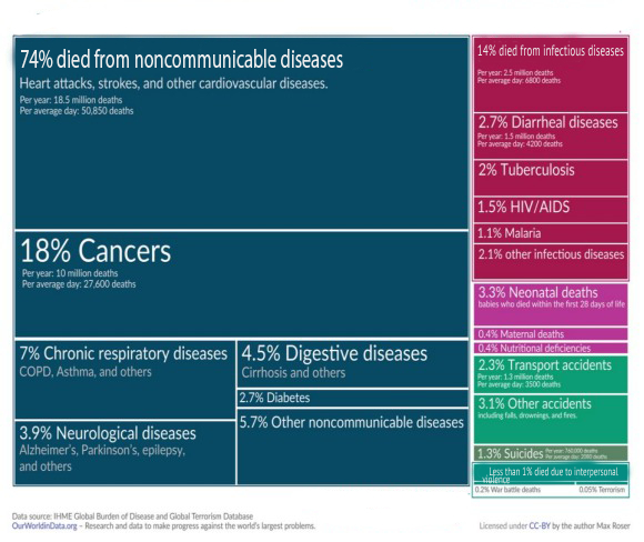
As per IHME Global Burden of Disease and Global Terrorism Database about 74% people in the world die from non- communicable diseases and percentage of these diseases is increasing every year. Each box represents one cause, and its size is proportional to the number of deaths it caused. The most common causes of death globally — shown in blue — were from ‘non-communicable diseases’ This includes cardiovascular diseases, cancer, and chronic respiratory diseases. Heart diseases were the most common cause, responsible for a third of all deaths globally. Cancers were in second, causing almost one-in-five deaths. Taken together, heart diseases and cancers are the cause of every second death. These diseases tend to develop gradually over time and aren’t infectious themselves. Thus, if we can catch these diseases in time the medical science is so developed that it can manage these diseases thus reducing risk of death and increasing the life expectancy.

At present the diagnosis is done by doctors looking at pictures of MRI, CT scan, X ray, etc. of patients and then comparing with other pictures which they seen during training or practice. Thus, the diagnosis is dependent upon the memory of the doctor.As about 74% of death in the world occurs due to non-communicable diseases as heart diseases, Cancer, respiratory diseases, digestive diseases, neurological diseases, diabetes, etc. Due to advancement of medical science now it is possible to easily manage these diseases if diagnosed within time. As early detection results in reduction in cost and complexity of treatment.

We offer the power, speed and accuracy of our FDA approved AI to the doctors to detect, analyse and treat these diseases effectively and in timely manner.

How did we read CT scan in 1980 How do we read CT scan in 2024
4 images on 8x10 film Images reviewed on computer (no film)
30-40 scan slices per case 2000-4000 scan slices per case
Acquisition time per study was 40-50 minutes (10 sec scan slides and 60 sec per slice reconstruction time) Acquisition time per study is 10 seconds or less with real time reconstruction (50 images/sec)
Limited resolution studies High resolution studies

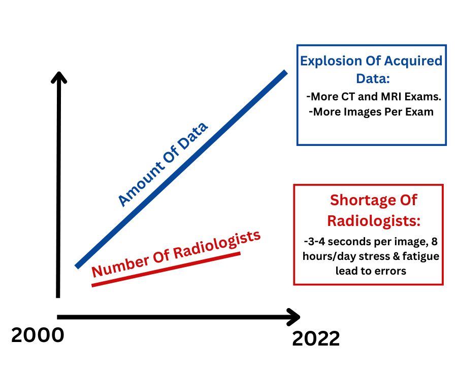
From graph it could be seen that as time passed the number of Radiologists has not kept pace with the increase of data. Thus the radiologist now have to read enormous amount of more data in same duty period of 8-12 hrs.

“A study was conducted to determine if increasing radiologist reading speed results in more misses and interpretation errors”. It was found that “Reading at the faster speed resulted in more major misses for 4 of the 5 radiologists. The total number of major misses for the 5 radiologists, when they reported at the faster speed, was 16 of 60 reported cases, versus 6 of the 60 reported cases at normal speed; P=0.032. The average interpretation error rate of major misses among the 5 radiologists reporting at the faster speed was 26.6% compared with 10% at normal speed.” The effect of Faster Reporting Speed for Imaging Studies on the Number of Misses and Interpretation Errors: A Pilot Study. Sokolovskaya E et al. J Am Coll Radiol. 2015 Jul; 12(7):683-8
In a similar study about reading the scans it was found that “ In the daily radiology practice, the rate of interpretation error is between 3% and 4%; however, of the radiology studies that contain abnormalities the error rate is even higher, averaging in the 30% range.” Also in this study “the majority of errors were the errors of underreading (42%), where the findings were simply missed.”
Fool me Twice: Delayed Diagnoses in Radiology with Emphasis on Perpetual Errors.
Kim YW, Mansfield LT, AJR 2014; 202:456-470

"Missed findings rather than misinterpretations of detected abnormalities were the most common reason for abdominopelvic CT report addenda. Awareness of the most common misses by anatomic location may help guide quality assurance initiatives. A wide variety of contributing factors were identified. Informatics and workflow optimization may be warranted to facilitate radiologists' access to all available patient-related data, as well as communication with other physicians, and thereby help reduce diagnostic errors."
Diagnostic errors in abdominopelvic CT interpretation: characterization based on report addenda
Andrew B. Rosenkrantz, Neil K. Bansal Abdom Radiol (2016)
41:1793-1799

How AI can help the doctors

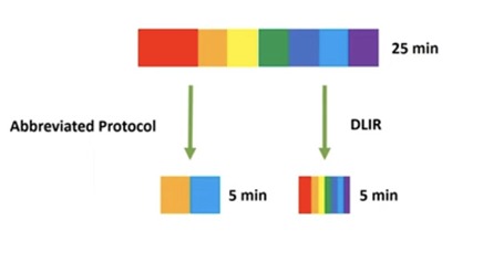
1) Deep Learning Image Reconstruction (DLR)

Deep learnings algorithm could be deployed for CT, MRI and PET scan image reconstruction. In this less time is required for scanning thus reducing chances movement of patient, less exposure to radiation and higher resolution, better contrast and less noise.

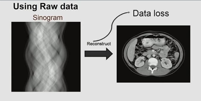
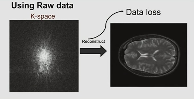
2) AI doesn’t require to see picture as it can directly read sonogram and K space waves

loss of data and noise and it doesn’t require proper contrast to read the scan picture.

3) AI can detect things which human eye can’t see.

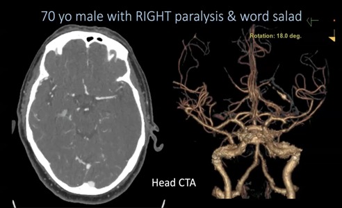
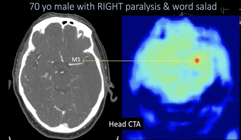
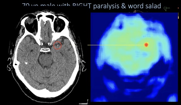
a) 70 yo male with Right paralysis & word salad head CTA (large surface occlusion model ) was normal and no defect was found, however the AI detected something.

then non contrast head CTA was checked and thrombus was found

b) in an 80 Yo female , usually 2mm slices are sent to radiologist but AI and examine even thinner slices as 0.6 mm and below. In one such case occlusion was found in thinner slice by AI which would have been missed by radiologist

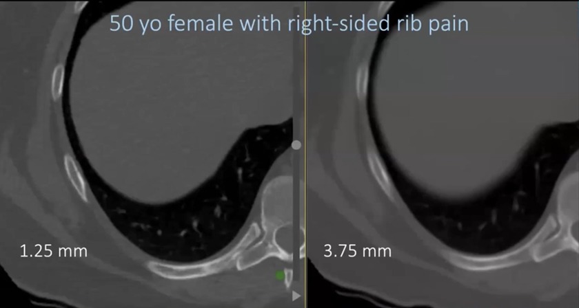
c) 50 yo female with rib fracture was exposed

4) AI helps doctors find unexpected findings

AI is different than humans, it does not look to images like we human do. we can find different things than what we were looking for as it looks at images differently (courtesy Radiology Partners)

a) AI is not biased by history

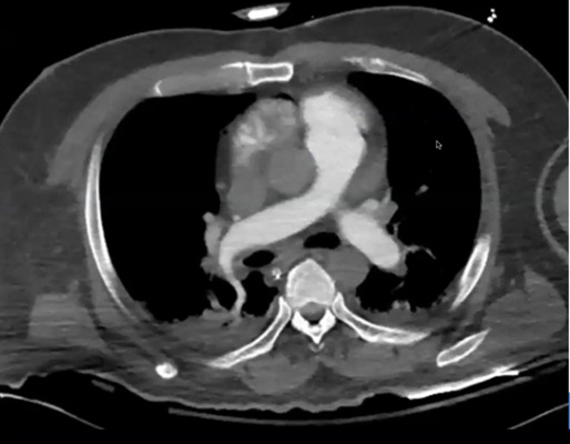
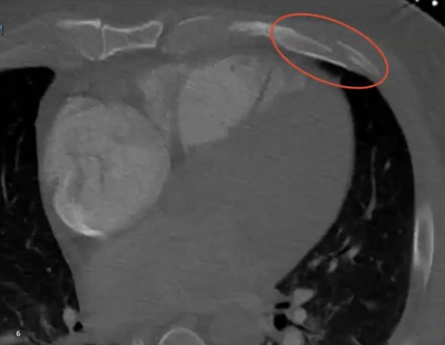
50 year old male with suspected PE, the Radiologist was searching in abdomen cavity but the AI found rib fracture

b) AI is not biased by satisfaction of search

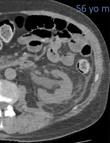
56 yo male with left flank pain , cause kidney hydrogenation, Radiologist missed tiny foci of free gas but AI caught it

c) AI is not biased by distracting pathology

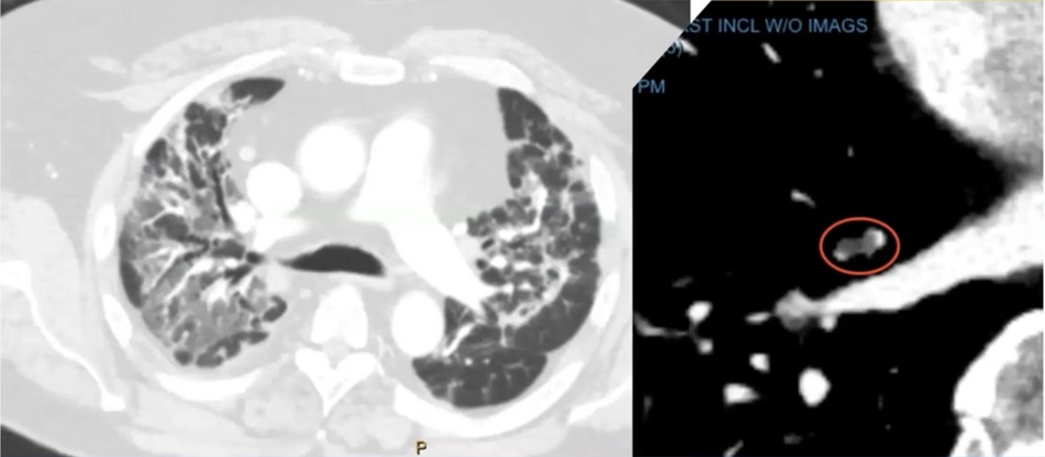
52 yo male with shortness of breadth during delta wave of covid. Radiologist was concentrated on lungs congestion but AI caught very small pulmonary emboli. (so small that missed by human eye)

d) AI is not biased by exam selection

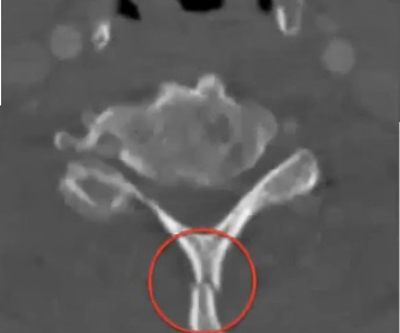
66 yo male with AMS (stroke) CT scan of brain , vessel occlusion no dissection was reported by Radiologist but AI caught cervical fracture.

e) AI is not biased by uncommon knowledge

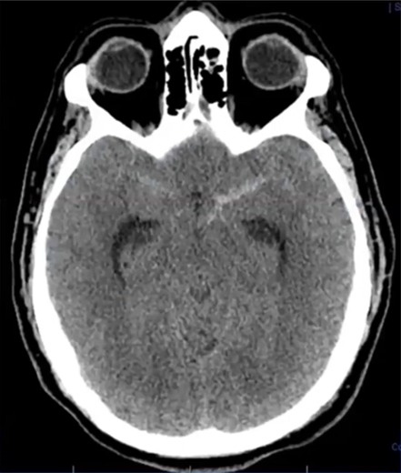
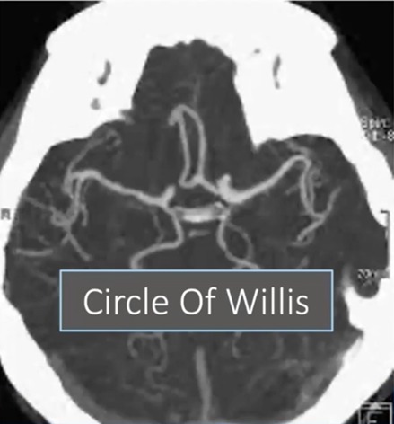
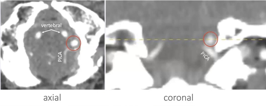
Head CT 47 yo male with SAH and Headache most common place for aneurism is circle of willis but it was not there. AI found aneurism in skull base.

5) AI helps Radiologist find subtle findings

AI helps Radiologist find subtle findings
blue2

Most of the findings done by AI is subtle findings as normal findings in routine are done by Radiologist, it is defects at unusual places where AI is required to help Radiologist. Thus, AI should Act as an analyst and Radiologist as consultant.

4) Cancer detection

in the realm of cancer early detection saves lives.

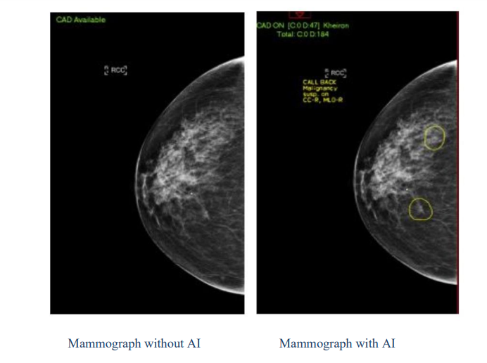
a) breast cancer

AI-Supported Mammogram Reading Detects 12% More Cancers

This research was published in the August 2023 issue of The Lancet Oncology.
“Artificial intelligence-supported screen reading versus standard double reading in the Mammography Screening with Artificial Intelligence trial (MASAI): a clinical safety analysis of a randomized, controlled, non-inferiority, single-blinded, screening accuracy study.”
Published in Aug 2023, there was an increase in 12% detection of Breast Cancer and 44.3 % reduction in screen reading workload.

B) Similarly for Lung cancer,

Experimental and clinical trial results demonstrate that deep learning techniques can be superior to trained radiologists. Deep learning is expected to effectively improve lung nodule segmentation, detection, and classification. With this powerful tool, radiologists can interpret images more accurately. Deep learning algorithm has shown great potential in a series of tasks in the radiology department and has solved many medical problems.

AI can be used to detect other Cancer and Brain Aneurysm

Conclusion

AI cannot pick everything but a team of AI and Radiologist can pick everything. This team which we are offering.

Future prospects

1) Finding Heart Cardiomegaly, i.e. detecting Heart attack long before they happen.

2) Finding Aortic Aneurysm, and brain aneurysm

3) Using Natural Language Processing to generate report. The AI detects each doctor’s pattern of writing report and then helps to write the complete report thus saving valuable time of Radiologist.